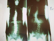
A little background. The brain and spinal cord are covered by a thin, protective sheath (or membrane) called the dura. It is extremely thin and delicate and it allows cerebrospinal fluid (CSF) to pass in and out, through and between as the CSF bathes not only the spinal cord but also the brain, keeping it happy and moist and in the brain's case, buoyant (which I will address the importance of later). Appropriate levels of CSF keep everything happy and in a state of homeostasis (remember that word from 7th grade science class?!?!) When you have too much fluid in the brain, you get something called hydrocephalus and often times a shunt is placed in the brain to drain out the excess CSF. In other cases where one doesn't have enough CSF, you get what is called "intracranial hypotension" (ICH, basically low blood pressure in the brain). The 3 most characteristic imaging features of ICH are (1) enhancement of the pachymeninges, or dura; (2) downward displacement of the brain ('brain sagging') and (3) enlarged pituitary. And lucky me, I happen to have all 3.
PHOTO #1: My brain MRI, which shows a thin white chalky outline especially in the front and top portion; the posterior portion is little more subtle but there nonetheless. You can also quite easily see something called 'brain sagging,' in which the most inferior posterior portion of my brain sort of bulges a bit.
PHOTO #2: Front and center you will notice a large dark spot (sorta look like a duck, LOL). At about 2 o'clock, you will notice a large pea-sized thing (that's my pituitary). It's apparently enlarged and should be about 1/4 of the size it is now.
PHOTO #3: This image clearly illustrates all of the white (that should NOT be present) in and around my brain, including the sulci (the 'valleys' and little nooks and crannies of the brain). The brain on MRI should be gray and black and the only white part should really just be the skills, the thick white outermost part.
PHOTO #4: The posterior aspect (looking at the inside of my brain as if you were standing behind me) illustrates quite a few things, all of which are indicative of a CSF leak. First, it again shows all of that white chalky outlining in my brain, and it even illustrates the sections of my brain (L, R and Cerebellum) quite easily because the lack of CSF literally 'outlines' my brain. Second, it shows 'brain sagging,' as you can very easily see that my cerebellum is basically sitting on the base of my skull, bumping up against the bony skull. (OUCH! PAIN!) Third, it also shows the dural enhancement surrounding my brain, very clearly in the top and upper side portions.
All of these images show what my neurosurgeon calls a "textbook CSF leak," where the brain is literally telling the doctors "Hey there's not enough fluid in here and we're not happy." These imaging findings cause a great deal of pain, which is why my head is KILLING me, the base of my neck and shoulders BURN, my eyesight is blurry (because the lack of fluid causes inflammation of the meninges and dura, which press on the optic nerve), whooshing in my ears (like I'm in a tunnel or a fan is blowing directly into my ears), and a few other things.
Looking at me, one would think "She looks ok to me" as there is nothing blatantly obvious - I'm not missing any limbs or getting around in a wheelchair. I can carry on a conversation. I can drive a car and do the laundry. It makes for a difficult explanation when on the outside I look fine but on the inside I'm a mess. My brain. My spine. My everything. This surgery HAS to be a success. There is just no room for anything BUT success....and failure is not an option. I hope my next blog entry is one where I am feeling good and doing well. I hope.
To read more about Low Pressure CSF headaches, click on the links below:


.JPG)
.JPG)

No comments:
Post a Comment